


Health sciences gains cutting-edge dissection technology
Posted on November 27, 2023
We place our future in their hands
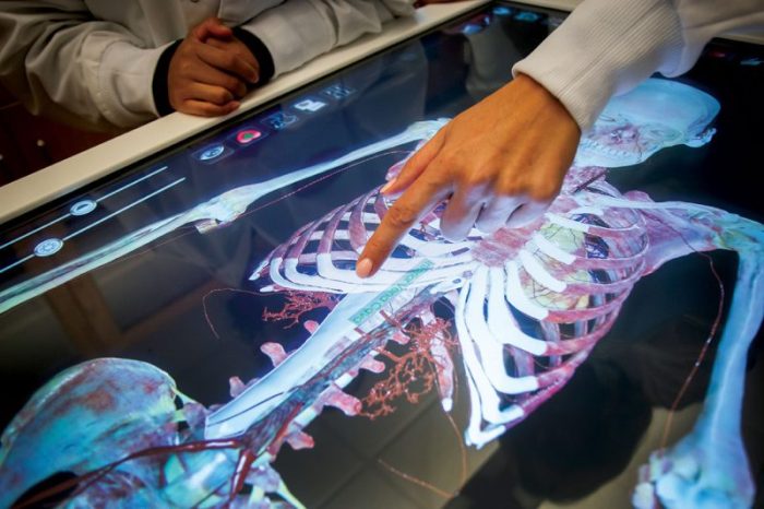
This fall, Mount Mary health sciences gained access to state-of-the-art technology with the arrival of a new Anatomage table. The table provides realistic anatomical modeling via 3D visualization technology. Using body scans from real cadavers, students can explore systems within the human body and interact with virtual anatomical models. Faculty can also customize lessons for the specific needs of their students, allowing for a diverse range of use across multiple programs within the Schools of Nursing, Arts and Sciences, Graduate Health and Professional programs.
The Anatomage table will provide students with a deeper understanding of complex bodily systems in ways that far surpass textbooks. The table provides a safe and ethical environment in which students can interact with biological specimens in a way previously only available through access to cadaver labs. And while the experience offers similarities to real-life dissection, it reinforces an even deeper understanding by allowing a user to repeat and review a process.
Since its arrival in August, the new Anatomage table has already been put into use by nursing, occupational therapy, pre-med, biology, exercise science, counseling and dietetics classes.
“This is a fascinating tool that offers the opportunity to investigate what the human body looks like at a variety of levels, from skin to muscle; bones, organs and more,” shares Janine Bamberger, assistant professor of dietetics. She cites the option to view five different cadavers available in the system, helping students understand individual differences that they will eventually encounter in real world practice.
“We can now offer our dietetics students both macro and micro views as we study how food becomes part of, and functions in, healthy and diseased bodies,” she adds.
Kari Inda, Ph.D., professor and chair of the occupational therapy department is also quickly embracing this technology in her classes.
“Having the ability to see the entire human body through true to life 3D imaging is greatly beneficial.”
Dr. Kari Inda, professor and chair of the occupational therapy department
“Having the ability to see the entire human body through true to life 3D imaging is greatly beneficial,” she states. “Instead of looking at images in textbooks or on computer screens, we can see full-size actual body images that have been digitized. We can rotate the body, dissect it, remove parts in layers, and isolate key structures to focus on how these structures work in solo or with other adjacent structures to create movement.”
Inda states that being able to visualize the body in layers will help students understand and apply therapeutic treatment to their patients. The Anatomage table will let students simulate X-ray, MRI and CT imaging, providing a high-resolution view of injuries and diseases from the inside.
The table was funded through a grant from the Howard Hughes Medical Institute (HHMI). This advanced technology is not yet available at many educational institutions, making the Anatomage table an especially meaningful addition to the university, and one which will undoubtedly prove an invaluable resource for Mount Mary classes for years to come.